Perfectly Prepped: 7 Reasons to Open Interproximal Contacts in Preparation for Porcelain Veneers
Maximize tooth preservation with a minimally invasive approach to open interproximal contacts in preparation for porcelain veneers. Keep reading to learn when to open interproximal contacts to create long-lasting veneers while maintaining tooth structure and maximum functionality.

Interproximal contact - to break, or not break thru the contact, that is the question.
When Should You Choose to Open Interproximal Contacts?

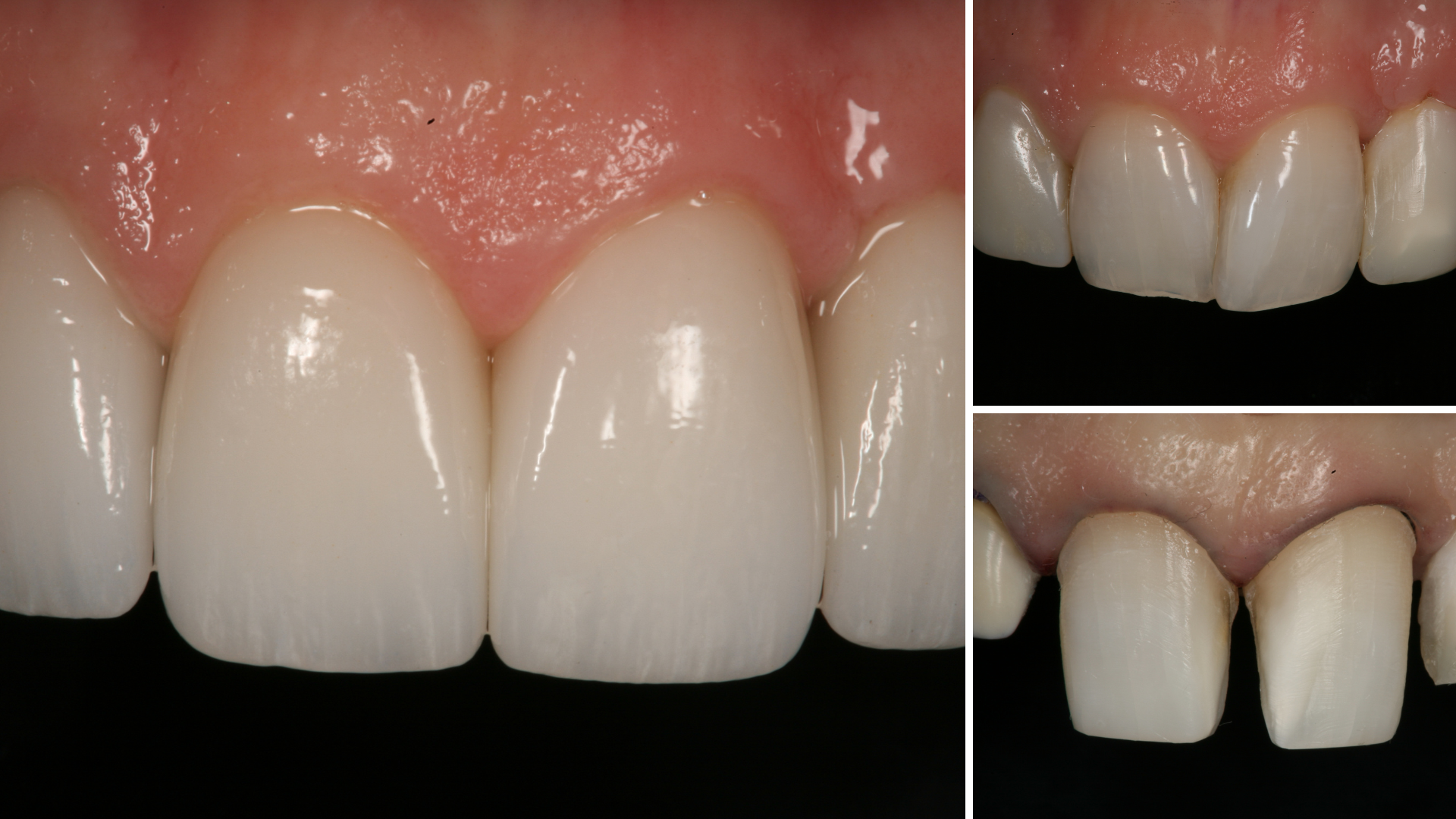
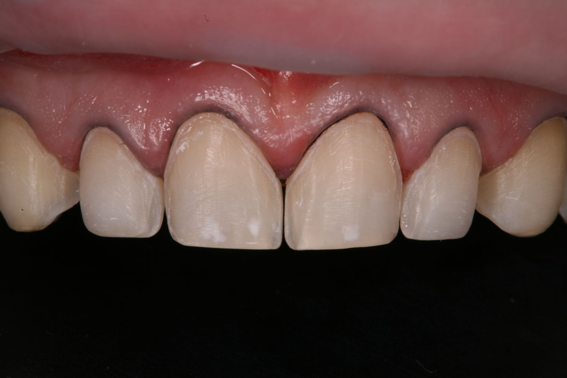
Note that the preparation margins are supragingival (above the gum line) and the proximal contacts of the natural teeth have been preserved by placing the margins facial to those contacts. While potentially more technically demanding for both clinician and laboratory technician, the desire to maintain tooth structure for the long-term health of the teeth is satisfied.
Also, and potentially equally important and relevant, the preparation is 100% in enamel, providing the best opportunity for optimal adhesion. Provided that the patient’s occlusion and parafunctional habits are managed appropriately, we would expect the porcelain veneers bonded exclusively to enamel, would maintain for many, many years, perhaps even decades.
There are times however, when for esthetic or functional reasons, the clinician needs to be more aggressive in their preparation design.
-
Closing diastemas
-
Heavy stain on natural teeth
-
Rotated teeth
-
Reduced enamel for bonding
-
Canted or angled midline
-
Laboratory dictates
-
Dark Teeth
1. Closing Diastemas
Figure 2
Figure 3
Essentially, the veneer material is cantilevered into the contact zone, and without the palatal support, the technician is not able to create proper tooth contour. If the veneer preparation margin extends to the lingual, more ideal contours of the veneer can be realized, providing for improved esthetics, and cleanliness of the veneer, as appropriate proximal contact contours and position can be created by the ceramist.
2. Overlapped & Rotated teeth

Figure 7
Figure 8
Figure 9
Figure 10
3. Canted or Angled Midline
Midline cants, or angled midlines are fairly common in the esthetic cases that we treat. The midline contact must be opened, and the proximal margins placed to the palatal, on cases with canted, or angled midlines. It is also important that all other angled or canted contacts be opened on teeth that are treated, or adjacent to the teeth being restored.
Opening the contacts allows the ceramist to 1) upright the contacts and make the contacts parallel to the face, and 2) create appropriate tooth proportions and symmetry between the teeth. The following illustrations (figure 11-17) and the image slides above demonstrate the restorative outcome with palatal margin placement.
Figure 11
Figure 12
Figure 13
Figure 14
Figure 16
Figure 17
4. Dark Teeth
Figure 18
Figure 19
Figure 20
Figure 21
Figure 22
Figure 23
Figure 24
Figure 25
For more on opaquing to bond dark teeth, check out our blog post, Blocking Out the Dark Tooth, or our complete course on DOT, Masking the Dark Tooth with Direct Resin Bonding.
5. Patients That Have Heavy Stains
The stain on porcelain is generally easily removed, however, when the staining occurs at the margin between the porcelain and tooth interface, those stains can be difficult, if not impossible, to completely remove.
6. Reduced Enamel
In cases where I am still attempting to be conservative in my preparation design, I might choose to do a facial “wrap” of the tooth to provide more retentive form to the veneer.
I like to tell the patients that in cases like these, the veneer will ‘hug’ the sides of the teeth. The interproximal wrap helps maintain the bonded restoration by increasing the amount of bonded tooth surface and by introducing retentive form in the restoration. The following clinical photographs demonstrate the ‘wrap veneer’ (Figure 26-28).
Figure 26
Figure 27
Figure 28
7. Laboratory Preference
Porcelain veneer restorations are an important tool in the cosmetic and restorative dentist’s tool box. Preparation designs however are exacting and precise to minimize tooth structure removal and provide biologically compatible, esthetic and functional restorations.
With conservative preparations, we are able to maintain as much enamel as possible, thereby providing the maximum long-term adhesion as possible for the porcelain veneers.
I feel strongly that positive collaboration between the dentist and the laboratory technician is critical for ultimate esthetics, and function, with all cosmetic cases, but most importantly with porcelain veneer cases due to the complexity of tooth preparation design.
Thanks for Reading!
Yours for better dentistry,
Dennis Hartlieb, DDS, AAACD
Share this page
Latest from our blog

CONNECT
Materials Included
Light Brown tints, Enamelize, Unfilled Resin Flexidiscs, Flexibuffs 1/2", #1 artist’s brush, Silicone Polishing Points, IPC Off Angle Short Titanium Coated Composite Instrument
Materials Needed, not Included
- Loupes
Follow along
You are Registered
-
Erosion and wear – the why and the how
-
Adding length to teeth – when is it safe
-
Opening VDO to compensate for lost tooth structure – where to begin
-
Records visit and key points you need to understand before you start
-
The smile – the 7 strategic points to consider when evaluating the smile
-
Anterior tooth shape, morphology
-
Clinical case review
-
Upper Putty matrix construction
-
Build lingual incisal wall with putty matrix #6 - #11/ Upper anteriors
-
Full contour build-up #6, #7, #8, #9, #10, #11, shape and polish/ Upper anteriors
-
Who – which patients are candidates
-
Why – explaining to patients the value of the prototype
-
How – step-by-step techniques to maximize predictability, efficiency and success
-
Getting to Yes: conversations with patients about esthetic and reconstructive dentistry
-
The ‘Smile Preview’ – techniques to show the possibilities
-
Lower Putty matrix construction
-
Build lingual incisal wall with putty matrix #22 - #27 / lower anteriors
-
Build-up #22 - #27, shape and polish / lower anteriors
-
Build-up lower occlusal posteriors
-
Demonstration of Smile Preview
Upcoming Virtual Workshops
-
01/22/2026
-
04/09/2026
-
06/11/2026
-
10/15/2026
-
12/10/2026
-
01/16/2026
-
02/13/2026
-
03/20/2026
-
04/10/2026
-
05/08/2026
-
06/05/2026
-
08/21/2026
-
10/09/2026
-
11/20/2026
-
12/10/2026
